
How 3D Imaging Enhances Root Canal Treatment & Oral Surgery
Dr. Labeeb
04 March 2025
Dental treatments require careful planning and accuracy. Root canals and oral surgeries involve working around delicate structures like nerves, blood vessels, and bone tissue. A mistake in these procedures can lead to pain, infections, or unsuccessful treatment.
For years, traditional dental X-rays have been the standard for diagnosing problems and planning procedures. However, X-rays provide only a two-dimensional image, making it difficult to see the full structure of teeth and bones. This limitation can lead to missed infections, hidden fractures, or improper placement of dental implants.
Understanding 3D Imaging in Dentistry
Dentists rely on imaging to assess the health of teeth, bones, and surrounding tissues. Traditional X-rays have served this purpose for many years, but they have limitations in depth and detail. 3D imaging provides a more comprehensive view, helping dentists detect hidden problems and make more informed treatment decisions.
What Is 3D Dental Imaging?
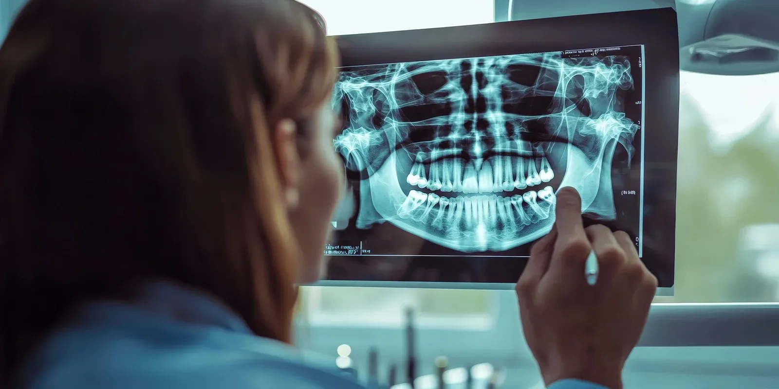
3D dental imaging, also known as Cone Beam Computed Tomography (CBCT), is an advanced imaging technique that produces a full three-dimensional scan of the mouth. Unlike traditional X-rays, which provide flat, two-dimensional images, CBCT captures the complete structure of teeth, jawbones, and soft tissues.
The process involves a specialized machine that rotates around the patient’s head, taking multiple images in seconds. These images are then compiled into a single 3D model, allowing dentists to examine the oral structures from multiple angles. This detailed view is particularly useful for diagnosing complex conditions and planning procedures with greater accuracy.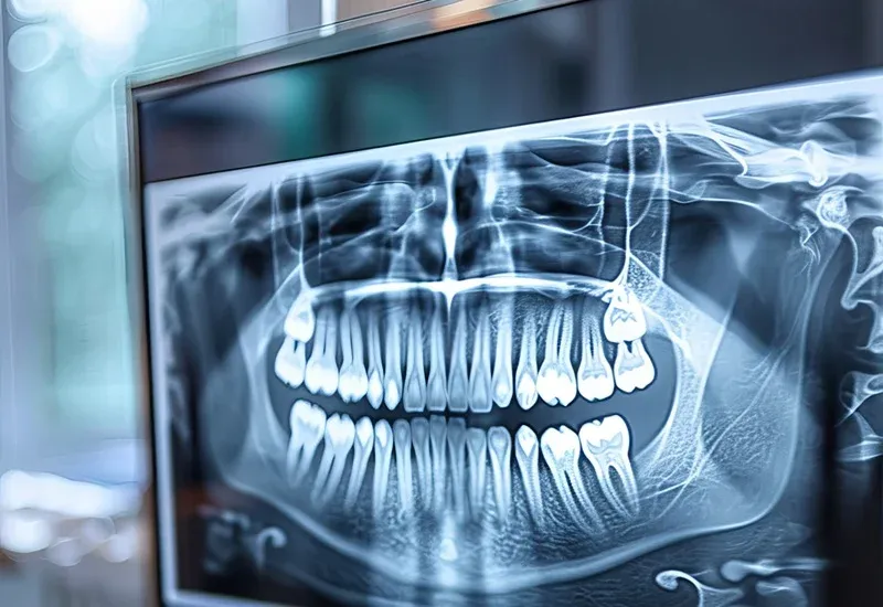
Why Dentists Prefer 3D Over Traditional X-Rays
While traditional X-rays remain useful for detecting cavities and routine dental assessments, they do not provide the level of detail needed for complex procedures like root canals, dental implants, or jaw surgeries.
CBCT offers several advantages over traditional X-rays:
- More Accurate Diagnosis – It reveals small fractures, hidden infections, and abnormalities that standard X-rays might miss.
- Better Treatment Planning – Dentists can measure bone density, locate nerves, and plan surgeries with precision.
- Improved Patient Safety – The ability to visualize critical structures helps prevent complications during surgery.
With 3D imaging, dental professionals can work with greater confidence, leading to improved patient outcomes.
3D Imaging in Root Canal Treatment
Root canals are delicate procedures that require precision. The goal is to remove infected or damaged pulp while preserving as much healthy tooth structure as possible. Traditional X-rays may not show all root canal pathways or detect infections deep inside the tooth.
Identifying Hidden Infections and Root Structures
Every tooth has multiple root canals, and their structure can be complex. Some roots have hidden or curved canals that are difficult to see on a standard X-ray. If a canal is missed during a root canal procedure, bacteria can remain inside the tooth, leading to reinfection.
CBCT imaging allows endodontists to see the entire internal structure of a tooth before treatment begins. This helps them locate all canals, identify small fractures, and assess the extent of infection. As a result, they can treat the problem more thoroughly and reduce the likelihood of complications.
Improving Precision in Root Canal Procedures
Successful root canal treatments depend on accuracy. Every infected canal must be thoroughly cleaned and sealed to prevent bacteria from spreading. With 3D imaging, dentists can:
- Determine the exact length and shape of root canals before starting treatment.
- Plan the safest and most efficient approach for removing infection.
- Ensure that all infected tissue is removed without damaging surrounding structures.
- This level of precision leads to more successful root canal treatments and fewer cases of retreatment.
Minimizing Treatment Discomfort
Many patients experience anxiety about root canals because of past experiences with discomfort. CBCT imaging helps reduce treatment time by allowing the dentist to work more efficiently. Because the entire tooth structure is visible in 3D, the dentist can avoid unnecessary drilling and make fewer adjustments during the procedure. This results in a more comfortable experience for the patient and a faster recovery.
3D Imaging in Oral Surgery
Oral surgery requires careful planning to ensure that procedures are safe and effective. Whether extracting a tooth or placing an implant, surgeons need to see all structures clearly to avoid complications. 3D imaging has made oral surgeries more predictable and less invasive.
Pre-Surgical Planning for Dental Extractions
Tooth extractions are not always straightforward. Some teeth have curved roots or are positioned close to nerves. Without a detailed image, there is a higher risk of complications, such as nerve damage or excessive bleeding.
With CBCT imaging, oral surgeons can assess the exact position of a tooth before extraction. They can determine the best approach to remove it while minimizing trauma to surrounding tissues. This reduces the risk of complications and promotes faster healing.
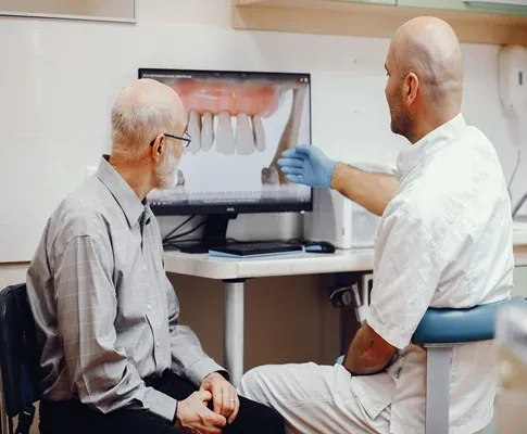
Enhancing Dental Implant Placement
Dental implants require precise placement to ensure long-term stability. The implant must be positioned in an area with enough bone support and away from sensitive structures like nerves or sinuses.
CBCT scans help oral surgeons plan implant placement with high accuracy. The 3D model allows them to measure bone density, assess the ideal implant angle, and simulate the procedure before surgery. This improves the success rate of implants and reduces healing time.
Reducing Surgical Risks
Oral surgeries involve working near delicate structures such as blood vessels and nerves. A miscalculation can lead to serious complications. CBCT scans allow oral surgeons to map out a procedure in advance, reducing the likelihood of unexpected issues during surgery. By knowing exactly where to operate, surgeons can work with greater confidence and precision.
The Role of 3D Imaging in Complex Dental Cases
Some dental conditions require advanced planning and multiple procedures. CBCT imaging is especially valuable in cases that involve jaw misalignment, bone grafting, or temporomandibular joint (TMJ) disorders.
Diagnosing TMJ Disorders
The temporomandibular joint (TMJ) controls jaw movement and plays a key role in chewing and speaking. TMJ disorders can cause pain, difficulty opening the mouth, and headaches. Diagnosing these conditions can be challenging because they involve soft tissues as well as bones.
3D imaging provides a detailed view of the jaw joint, allowing dentists to identify structural issues. They can detect problems such as joint misalignment, bone degeneration, or inflammation. With this information, they can develop a treatment plan that targets the root cause of the problem.
Need help?
Contact usAssessing Bone Grafts for Implants
Some patients do not have enough bone to support a dental implant. In these cases, a bone graft may be necessary to strengthen the jaw.
CBCT scans help dentists determine whether a patient needs a bone graft. They can measure bone thickness, identify weak areas, and plan graft placement with accuracy. This improves the chances of a successful implant and reduces the risk of failure.
Planning for Corrective Jaw Surgery
Corrective jaw surgery is used to fix problems with bite alignment, facial structure, or breathing difficulties. These procedures require careful planning because they involve repositioning the jawbone.
CBCT imaging allows surgeons to assess the jaw structure in three dimensions. They can visualize how the bones fit together and determine the best way to correct misalignment. This results in a safer, more predictable surgery with better long-term outcomes.
Conclusion
3D imaging has changed the way dentists and oral surgeons approach treatment. By providing clear, detailed images of the teeth and jaw, it improves accuracy, reduces risks, and enhances patient comfort. Whether diagnosing an infection, planning a root canal, or preparing for surgery, CBCT technology helps dental professionals work with greater confidence.
For patients, this means more precise treatments, shorter recovery times, and better overall results. As technology continues to advance, 3D imaging will remain an essential tool in modern dentistry, ensuring safer and more effective care for years to come.
Share This:
Disclaimer
*This media/content or any other on this website does not prescribe, recommend, or prevent any treatment or procedure. Therefore, we highly recommend that you get the advice of a qualified dentist or other medical practitioners regarding your specific dental condition. *