

Innovative Dental Solutions
Contact Us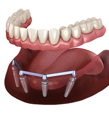
READY TO GET SERVICE?
GENERAL AND FAMILY DENTISTRY
SEDATION DENTISTRY
PEDIATRIC DENTISTRY
COSMETIC DENTISTRY
ORTHODONTICS
ADVANCED DENTAL TECHNOLOGY
Procure Healthy Smiles
You can experience the dentistry that goes beyond the basics with Studio City Dental Group.

3D Dental Imaging Benefits: What X-rays Can Miss
Dr. Melody Khonsari
November 20, 2026
You have a question that comes up when a dentist wants a clearer view of a tooth, a root, or the bone around it, what can 3D dental imaging show that standard X-rays may not? At Studio City Dental Group, Dr. Melody Khonsari uses imaging as part of a larger exam, so the goal is to see the structures that matter for diagnosis and planning, not to rely on one picture alone.
The Digital X-rays & 3D Wellness Scans service gives the team a way to look beyond a flat image when a case needs more depth. Standard X-rays still have a place, and 3D imaging has its own role, depending on what the exam shows and what the dentist is trying to understand.
For many adults, the value is in seeing fine detail around roots, bone, and nearby anatomy before a procedure starts. That detail can help the dentist review a problem tooth from more than one angle and decide whether the 2D view is enough or a three-dimensional scan would add useful information.
What 3D imaging can add
A 3D scan creates a layered view of the mouth and jaw, so the dentist can study a tooth from the front, side, and depth instead of only a single flattened image. That matters when the question is not just where a tooth sits, but how its root shapes, the surrounding bone, and nearby anatomy line up in space.
In everyday care, that extra view can help with root canal planning, implant evaluation, wisdom teeth assessment, and some surgical cases. The image can also show small areas where bone has changed, a hidden root canal path, or the position of structures that sit close to treatment areas.
During an exam at Studio City Dental Group, Dr. Melody Khonsari looks at your symptoms, the tooth or area of concern, and any previous imaging before deciding what kind of scan is useful. The team explains what the scan is meant to show, so the visit stays focused on your specific diagnosis rather than a routine image for its own sake.
3D dental imaging benefits when detail matters
The main 3D dental imaging benefits come from seeing anatomy in full depth. A standard X-ray flattens structures together, which can hide overlap between teeth, roots, bone, and sinuses. A 3D scan separates those structures so the dentist can study them more clearly.
That added detail is often helpful when the exam suggests a complex problem. It may show the shape of a root tip, the extent of bone loss, the path of a canal, or the location of a tooth that has not fully come in. It can also show how close a planned treatment area sits to nerves, the sinus floor, or adjacent tooth roots.
The scan does not replace the exam. It adds information when the dentist needs more than a flat image, and it is used with other findings such as symptoms, probing, movement, bite changes, and the look of the tooth itself.
Where standard X-rays still help
Standard dental X-rays remain useful in many routine situations. They can show cavities between teeth, changes around the root tip, bone levels, and the fit of restorations in a straightforward way. For many checkups, they provide enough information for the dentist to monitor oral health.
Because they use less radiation than a 3D scan and answer many routine questions, they are often a practical first look. A dentist may begin with regular X-rays and only move to a three-dimensional image if the findings are not clear enough or if the treatment plan calls for more precise anatomy.
That approach is one reason imaging is tailored to the case. A scan is selected for a reason, and the choice depends on what question needs answering at that visit.
- Routine cavity checks often start with standard X-rays.
- A root canal case may need depth to trace canal anatomy.
- Implant planning may need bone height and width measured in 3D.
- Wisdom tooth positions can be clearer when roots are seen from multiple angles.
- Some jaw or sinus concerns need a broader view than a flat film can provide.
What a 3D scan may show that a 2D image can miss
Three-dimensional imaging can reveal details that are easy to miss when structures overlap. For example, one tooth root may sit behind another on a standard image, or a bone contour may look different once the scan is viewed from the side. That can change how the dentist interprets the area.
It can also show the actual relationship between a tooth and nearby structures. In the upper back teeth, that includes the sinus. In the lower jaw, that may include the nerve canal. In both areas, the benefit is less about making a diagnosis from one image alone and more about mapping the area accurately before treatment.
The American Dental Association notes that dental imaging is used to help evaluate teeth, roots, and surrounding structures, and that the choice of image depends on the clinical need. You can read more on the ADA's overview of dental X-rays.
For some patients, the scan also helps explain why a symptom has been hard to localize. Pain can refer to a nearby tooth, a cracked root can be hidden below the gumline, or bone changes can sit in an area that does not show well on a small film. The scan does not tell the whole story by itself, but it can uncover the part that was missing.
When a dentist may choose 3D imaging
A 3D scan is more likely when the mouth needs a closer, more spatial view. That is true in treatment planning and in diagnosis, especially when a routine image leaves unanswered questions.
Common examples include:
- planning a dental implant and measuring bone around the site
- checking the path of a root canal or a retreatment case
- looking at impacted or angled wisdom teeth
- reviewing a jaw area before oral surgery
- evaluating bone loss or anatomy around the sinuses
For patients who want a broader overview of how this technology fits into care, the earlier post, What 3D Dental Imaging Shows That a Standard X-Ray Can Miss, looks at a common misconception about these scans. It pairs well with this article, which looks more closely at when that extra detail helps.
One practical example is implant planning. A 3D image can show the width of bone, the height of bone, and nearby anatomy in a way a flat image cannot. That information helps the dentist review whether the area is shaped in a way that supports the planned treatment, and it is one reason the Dental Implants page is often linked with imaging discussions.
What the visit usually involves
The scan itself is typically quick, and the chair time is usually short. You stand or sit still while the machine moves around the head, then the images are reviewed by the dental team. The important part is not the scan alone, but how the dentist interprets it with the rest of the exam.
At Studio City Dental Group, that review may include prior X-rays, a visual check, and the reason you came in. Dr. Melody Khonsari can then compare the findings and decide whether the 3D image answered the question or whether another type of imaging is more appropriate for the case.
If you want to read more about how regular imaging fits into routine care, the earlier post Why Do I Need Dental X-Rays gives a useful baseline for why dentists use imaging at all. This article goes one step further and focuses on what changes when the view becomes three-dimensional.
A dentist may choose 3D imaging more often for complex treatment planning, while still relying on standard X-rays for everyday monitoring. The right choice depends on the clinical question, not on using the most detailed image every time.
“A 3D scan adds depth to the exam, so the dentist can see relationships that a flat image may hide.”
FAQ
Still have a Question? Contact Us
No. Regular X-rays still help with many routine checks, especially when the question is straightforward. A 3D scan is used when the dentist needs more depth or a wider view of the area.
It can show the exact position of roots, bone contours, and nearby structures such as nerves or sinus spaces. It also makes overlapping anatomy easier to sort out.
Not always, but implant planning often benefits from the extra measurements a scan can provide. The dentist decides based on the bone, the location, and the treatment plan.
It can, especially when pain is hard to pinpoint or when a problem sits below the surface. The scan gives more spatial detail, which may help the dentist look for hidden causes.
The decision comes from the exam, your symptoms, and the area being studied. Imaging is chosen to answer a specific clinical question, not as a routine step for every visit.
Conclusion
3D dental imaging gives the dentist a deeper look at teeth, roots, bone, and nearby structures, which can make certain diagnoses and treatment plans clearer. Standard X-rays still matter, but they do not show depth the same way, so the best image depends on the question being asked.
At Studio City Dental Group, Dr. Melody Khonsari uses imaging as part of a careful exam, which helps the team decide when a flat film is enough and when a 3D scan adds useful detail. The next post in this series looks at how a digital or 3D dental X-ray works and what it shows.
Our sister practice, One Dental Studio City, explains this service on its website.
To talk through your imaging options with Studio City Dental Group, contact the practice and schedule a visit with Dr. Melody Khonsari.
More in this series
Share This:
Disclaimer
This media/content or any other on this website does not prescribe, recommend, or prevent any treatment or procedure. Therefore, we highly recommend that you get the advice of a qualified dentist or other medical practitioners regarding your specific dental condition.